This advanced module is presented to the 3rd year students later in their clerkship, after they’ve had training in basic suturing, and have had a chance to scrub into surgery.
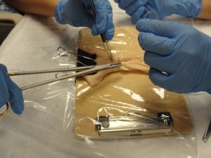
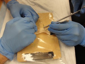
The students work in pairs (Surgeon, and Assistant) to close two lacerations in intestines (cow or pig, depending on availability).

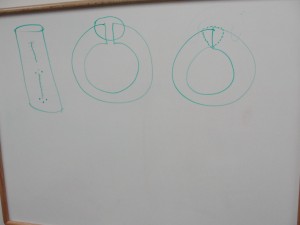
Before beginning their repair, they are given brief instructions on how to close these lacerations in two layers. First, the outer two-thirds (not perforating the mucosa), followed by an imbricating layer to bury the first layer, creating a water-tight closure.
Grease board drawings illustrate the closure technique.Each intestine has two short, longitudinal lacerations. This enables each student to close one as a Surgeon, and to assist in the closure of one as an Assistant. By keeping the lacerations short, it doesn’t take a long time for them to close the wounds.
Because of time constraints (one hour), the students are asked to use running sutures rather than interrupted (although they are told that in real patients, these sutures are frequently interrupted.)
As they are suturing the wounds, faculty circulate among the teams, giving guidance, insights, and correcting poor technique.

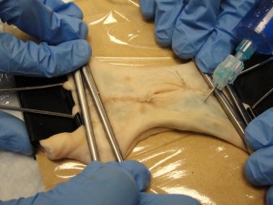
After both lacerations are closed, blue dye is injected into the lumen to test the water tightness of each student’s repair.

This is an imperfect model. The intestine is too thin and dead for a high fidelity representation of the human intestine. It often has minor imperfections that leak when tested.
Further, it is infrequent that an intestinal laceration would be repaired using this technique. But the point is not to train the M3s to be intestinal surgeons…it is to give them experience in suturing and assisting in a controlled environment, with careful teaching from knowledgeable faculty.

